Anti-TAB2: Mouse TAB2 Antibody |
 |
BACKGROUND Transforming growth factor-β-activated kinase 1 (TAK1), a member of the MAP3K family, was initially identified as a kinase in transforming growth factor β signaling. Recent evidence has emerged indicating that TAK1 is involved in various signaling pathways, including IL-1, IL-18, TNF, and receptor activator of NF-κB ligand (RANKL). Members of the TNF receptor-associated factor (TRAF) family of adaptor proteins are involved in coupling activated TNF and IL-1 receptors to NF-κB and MAPKs pathways. Although TRAF proteins have no known enzymatic activity, recent evidence indicates that they act as E3s through their N-terminal really interesting new gene (RING) domain. RING-type E3s contain a series of Cys and His residues distinctly separated to constitute a novel zinc-binding domain. TRAF6 together with the heterodimeric E2 complex (Ubc13/Uev1A) facilitates the synthesis of unique Lys63-linked polyubiquitin chains on itself, rather than the conventional Lys48-linked polyubiquitin chains that target proteins for degradation. This unique Lys63-linked polyubiquitin chain likely provides a scaffold to recruit downstream effector molecules to activate various signaling components.1
TAK1 binding protein 2 (TAB2) is one of three proteins that are constitutively bound to TAK1. TAK1 binding protein 1 (TAB1) acts as the activation subunit of the TAK1 complex, aiding in the autophosphorylation of TAK1; while TAB2 and the closely related protein, TAB3, are adaptors of TAK1 that recruit TAK1 to a TNF receptor signaling complex and facilitate the assembly of an active TAK1 complex. Following IL-1, TNF, or LPS stimulation, two possible TAK1 complexes exist, one which consists of TAB2-TAK1-TAB1 and another that consists of TAB3-TAK1-TAB1, which causes the activation of MAPKs and NF-κB pathways.2 TAB2 contains 3 conserved domains: a CUE domain that directly binds to ubiquitin, a coiled-coil domain involved in the interaction with TAK1, and a zinc finger domain which is involved in polyubiquitin binding. TAB2 and TAB3 bind the polyubiquitin chain and TAK1, thereby bringing TAK1 in close proximity to the polyubiquitinated proteins RIP1 and TRAF and the IKK complex. TAB2 and TAB3 redundantly mediate activation of TAK1. It was shown that TAB2 is not essential for IL-1 signaling pathways due to the presence of TAB3, which can play a compensatory role. However, TAB2 single knockout mice are embryonic lethal at E13.5, indicating that TAB2 has some unique functions that cannot be compensated by TAB3.3 Moreover, TAB2-deficient fibroblasts displayed a significantly prolonged activation of TAK1 compared with wild type control cells. This suggests that TAB2 mediates deactivation of TAK1. Furthermore, it was demonstrated that a TAK1 negative regulator, protein phosphatase 6 (PP6), was recruited to TAK1 complex in wild type but not in TAB2-deficient fibroblasts. Additionally, it was shown that both PP6 and TAB2 interacted with the polyubiquitin chains and this interaction mediated the assembly with TAK1. Thus, TAB2 not only activates TAK1 but also plays an essential role in the deactivation of TAK1 by recruiting PP6 through a polyubiquitin chaindependent mechanism.4 Recently, it was shown that TAB2 could directly interact with NLK and function as a scaffold protein to facilitate the interaction between TAK1 and NLK. Knocking down TAB2 using small interfering RNA abolished the interaction of TAK1 with NLK in mammalian cells. Moreover, Wnt3a stimulation led to an increase in the interaction of TAB2 with NLK and the formation of a TAK1•TAB2•NLK complex, suggesting that this TAK1-TAB2-NLK pathway may constitute a negative feedback mechanism for canonical Wnt signaling.5
TAK1 binding protein 2 (TAB2) is one of three proteins that are constitutively bound to TAK1. TAK1 binding protein 1 (TAB1) acts as the activation subunit of the TAK1 complex, aiding in the autophosphorylation of TAK1; while TAB2 and the closely related protein, TAB3, are adaptors of TAK1 that recruit TAK1 to a TNF receptor signaling complex and facilitate the assembly of an active TAK1 complex. Following IL-1, TNF, or LPS stimulation, two possible TAK1 complexes exist, one which consists of TAB2-TAK1-TAB1 and another that consists of TAB3-TAK1-TAB1, which causes the activation of MAPKs and NF-κB pathways.2 TAB2 contains 3 conserved domains: a CUE domain that directly binds to ubiquitin, a coiled-coil domain involved in the interaction with TAK1, and a zinc finger domain which is involved in polyubiquitin binding. TAB2 and TAB3 bind the polyubiquitin chain and TAK1, thereby bringing TAK1 in close proximity to the polyubiquitinated proteins RIP1 and TRAF and the IKK complex. TAB2 and TAB3 redundantly mediate activation of TAK1. It was shown that TAB2 is not essential for IL-1 signaling pathways due to the presence of TAB3, which can play a compensatory role. However, TAB2 single knockout mice are embryonic lethal at E13.5, indicating that TAB2 has some unique functions that cannot be compensated by TAB3.3 Moreover, TAB2-deficient fibroblasts displayed a significantly prolonged activation of TAK1 compared with wild type control cells. This suggests that TAB2 mediates deactivation of TAK1. Furthermore, it was demonstrated that a TAK1 negative regulator, protein phosphatase 6 (PP6), was recruited to TAK1 complex in wild type but not in TAB2-deficient fibroblasts. Additionally, it was shown that both PP6 and TAB2 interacted with the polyubiquitin chains and this interaction mediated the assembly with TAK1. Thus, TAB2 not only activates TAK1 but also plays an essential role in the deactivation of TAK1 by recruiting PP6 through a polyubiquitin chaindependent mechanism.4 Recently, it was shown that TAB2 could directly interact with NLK and function as a scaffold protein to facilitate the interaction between TAK1 and NLK. Knocking down TAB2 using small interfering RNA abolished the interaction of TAK1 with NLK in mammalian cells. Moreover, Wnt3a stimulation led to an increase in the interaction of TAB2 with NLK and the formation of a TAK1•TAB2•NLK complex, suggesting that this TAK1-TAB2-NLK pathway may constitute a negative feedback mechanism for canonical Wnt signaling.5
REFERENCES
1. Landstrome, M.: Int. J. Biochem. cell Biol. 42:585-9, 2010
2. Besse, A. et al: J. Biol. Chem. 282:3918-28, 2007
3. Kulathu, Y. et al: Nature Struct. Mol. Biol. 16:1328-30, 2009
4. Broglie, P. et al: J. Biol. Chem. 285:2333-9, 2010
5. Li, M. et al: J. Biol. Chem. 285:13397-404, 2010
2. Besse, A. et al: J. Biol. Chem. 282:3918-28, 2007
3. Kulathu, Y. et al: Nature Struct. Mol. Biol. 16:1328-30, 2009
4. Broglie, P. et al: J. Biol. Chem. 285:2333-9, 2010
5. Li, M. et al: J. Biol. Chem. 285:13397-404, 2010
Products are for research use only. They are not intended for human, animal, or diagnostic applications.
Параметры
Cat.No.: | CP10297 |
Antigen: | Raised against recombinant human TAB2 fragments expressed in E. coli. |
Isotype: | Mouse IgG1 |
Species & predicted species cross- reactivity ( ): | Human, Mouse, Rat |
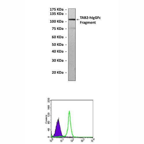
Applications & Suggested starting dilutions:* | WB 1:1000 IP 1:50 IHC n/d ICC n/d FACS 1:50 - 1:200 |
Predicted Molecular Weight of protein: | 80 kDa |
Specificity/Sensitivity: | Detects endogenous TAB2 proteins without cross-reactivity with other family members. |
Storage: | Store at -20°C, 4°C for frequent use. Avoid repeated freeze-thaw cycles. |
*Optimal working dilutions must be determined by end user.
Документы
Информация представлена исключительно в ознакомительных целях и ни при каких условиях не является публичной офертой


