

Anti-Myostatin: Mouse Myostatin Antibody |
 |
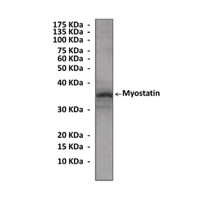
BACKGROUND Myostatin (MSTN) (initially termed growth and differentiation factor 8, GDF-8), a member of the transforming growth factor-beta (TGF-beta) superfamily, has been established as a regulator of development and growth in several vertebrates. During development and adulthood, MSTN is predominately expressed in skeletal muscle, though there have been reports of myostatin protein in cardiomyocytes and Purkinje fibers of the heart, as well as MSTN mRNA expression in the mammary gland.1 Just as with other members of the TGF-beta superfamily, MSTN is synthesized as a precursor protein. Removal of the signal sequence is followed by proteolytic cleavage at a specific tetrabasic processing site. There are numerous reports of the active MSTN protein in mammals ranging in size from 26 to 30 kDa. It has been demonstrated that post-translational modifications occur in vivo. However, recent research examining serum protein from MSTN deficient and wild-type adult mice has shown that a 12.5 kDa protein is the biological active MSTN form, not the peptide that is approximately 26 kDa.2
Myostatin was first defined as a negative regulator of muscle mass on the basis of a mouse model. Myostatin functions mainly to modulate myoblast proliferation and thus muscle mass and strength. Myostatin overexpression leads to an accumulation of myoblasts in the G0/G1 and G2 phases and thus are not available for differentiation into myotubes. It was further shown that myostatin exerts its effects by moderating myogenin and increasing the expression of p21 and p53, both of which are involved in cell-cycle control. In addition, a step past proliferation of myoblasts on the way to muscle fibers is their differentiation, which appears to be affected by myostatin as well. Moreover, Myostatin seems to have some other peripheral effects. It negatively regulates apoptosis in muscle cells. Variants of the MSTN gene are associated with muscle hypertrophy phenotypes in a range of mammalian species, most notably cattle, dogs and mice. The myostatin-null mouse model also provides insights into the physiological role of this protein. Besides its function in reducing sarcopenia, it appears that myostatin also regulates the structure and function of tendon tissues, as the stiffness of tendons is 14 times higher in myostatin-deficient mice than in their wild type controlsGrowth hormone might be a potential inhibitor of myostatin production. Interestingly, myostatin also has a significant effect on adipose tissue. It was reported that myostatin promotes adipogenesis and myostatin antibodies suppress adipogenesis using a marker of adipogenesis with pluripotent cells. It was also demonstrated that myostatin can determine the fate of multipotent stem cells, but not preadipocytes, and that this effect leads to adipocytes with increased insulin sensitivity. Knockout of myostatin can decrease body fat and promote resistance against developing atherosclerosis, or "hardening" of the arteries. Molecularly then, myostatin has favorable effects on the development of both skeletal muscle and adipose tissue.3 Myostatin and its negative effects on skeletal muscle mass have understandably captivated many biomedical, agricultural, and comparative biologists, since the gains in muscle mass associated with the myostatin null phenotype have never been reproduced by the administration of growth promoters regardless of species or mode of administration. The potential benefits of reproducing these effects in the clinic or in animal feed lots are obvious and cannot be overestimated. Relieving myostatin's restrictive effects on skeletal muscle growth and development could revolutionize the clinical treatment of different muscle growth disorders, including some muscular dystrophies, and has the potential to significantly enhance the production of meat animal products as well.4
Myostatin was first defined as a negative regulator of muscle mass on the basis of a mouse model. Myostatin functions mainly to modulate myoblast proliferation and thus muscle mass and strength. Myostatin overexpression leads to an accumulation of myoblasts in the G0/G1 and G2 phases and thus are not available for differentiation into myotubes. It was further shown that myostatin exerts its effects by moderating myogenin and increasing the expression of p21 and p53, both of which are involved in cell-cycle control. In addition, a step past proliferation of myoblasts on the way to muscle fibers is their differentiation, which appears to be affected by myostatin as well. Moreover, Myostatin seems to have some other peripheral effects. It negatively regulates apoptosis in muscle cells. Variants of the MSTN gene are associated with muscle hypertrophy phenotypes in a range of mammalian species, most notably cattle, dogs and mice. The myostatin-null mouse model also provides insights into the physiological role of this protein. Besides its function in reducing sarcopenia, it appears that myostatin also regulates the structure and function of tendon tissues, as the stiffness of tendons is 14 times higher in myostatin-deficient mice than in their wild type controlsGrowth hormone might be a potential inhibitor of myostatin production. Interestingly, myostatin also has a significant effect on adipose tissue. It was reported that myostatin promotes adipogenesis and myostatin antibodies suppress adipogenesis using a marker of adipogenesis with pluripotent cells. It was also demonstrated that myostatin can determine the fate of multipotent stem cells, but not preadipocytes, and that this effect leads to adipocytes with increased insulin sensitivity. Knockout of myostatin can decrease body fat and promote resistance against developing atherosclerosis, or "hardening" of the arteries. Molecularly then, myostatin has favorable effects on the development of both skeletal muscle and adipose tissue.3 Myostatin and its negative effects on skeletal muscle mass have understandably captivated many biomedical, agricultural, and comparative biologists, since the gains in muscle mass associated with the myostatin null phenotype have never been reproduced by the administration of growth promoters regardless of species or mode of administration. The potential benefits of reproducing these effects in the clinic or in animal feed lots are obvious and cannot be overestimated. Relieving myostatin's restrictive effects on skeletal muscle growth and development could revolutionize the clinical treatment of different muscle growth disorders, including some muscular dystrophies, and has the potential to significantly enhance the production of meat animal products as well.4
REFERENCES
1. McNally, E.M.: N. Engl. J. Med. 350:2642-4, 2004
2. Roberts, S.B. & Goetz, F.W.: Mol. Cell. Endocrinol. 210:9:20, 2003
3. Weiner, G. et al: Eukaryon 5:91-4, 2009
4. Santiago, C. et al: PLoS ONE 6:e16323, 2011
2. Roberts, S.B. & Goetz, F.W.: Mol. Cell. Endocrinol. 210:9:20, 2003
3. Weiner, G. et al: Eukaryon 5:91-4, 2009
4. Santiago, C. et al: PLoS ONE 6:e16323, 2011
Products are for research use only. They are not intended for human, animal, or diagnostic applications.
Параметры
Cat.No.: | CP10315 |
Antigen: | Raised against recombinant human myostatin fragments expressed in E. coli. |
Isotype: | Mouse IgG1 |
Species & predicted species cross- reactivity ( ): | Human, Mouse, Rat |
Applications & Suggested starting dilutions:* | WB 1:1000 IP n/d IHC n/d ICC n/d FACS n/d |
Predicted Molecular Weight of protein: | 12-30, 49-52 kDa |
Specificity/Sensitivity: | Detects endogenous myostatin proteins without cross-reactivity with other family members. |
Storage: | Store at -20°C, 4°C for frequent use. Avoid repeated freeze-thaw cycles. |
*Optimal working dilutions must be determined by end user.
Документы

Информация представлена исключительно в ознакомительных целях и ни при каких условиях не является публичной офертой


