Anti-PINCH1: Mouse PINCH1 Antibody |
 |
BACKGROUND PINCH1 (particularly interesting new cysteine-histidine rich protein 1) is an adaptor protein that plays an important role in regulating cell spreading, motility, epithelial-mesenchymal transition and matrix production. Structurally, PINCH1 contains a tandem array of five LIN11, Isl1 and MEC-3 (LIM) domains that are involved in mediating protein-protein interactions, and a short C-terminal tail that harbors a putative leucine-rich nuclear export signal (NES) and overlapping basic nuclear localization signal (NLS). PINCH1 is developmentally regulated and its expression is critical for proper cytoskeletal organization and extracellular matrix adhesion. Although PINCH1 has no catalytic abilities, the PIP (PINCH1–ILK–parvin) complex serves as a link between integrins and components of growth factor receptor kinase and GTPase signaling pathways. Furthermore, the PINCH1 proteins have also been shown to bind Nck adapter protein 2 (Nck2), Ras suppressor protein 1 (Rsu1), and thymosin β4. In concert with transmembrane integrin and growth factor receptors, the PINCH1/ILK/parvin complex enables cooperative signaling for the regulation of cell survival via the PI3K/PKB/Akt1 and Ras/MAPK cascades. It was shown that PINCH1 is able to modulate PKB/Akt activity in an ILK-dependent and an ILK-independent manner and moreover, that PINCH1 functions in the survival pathway both upstream of PKB/Akt as well as downstream or alternatively, independent of PKB/Akt. In addition, Akt1 can be directly dephosphorylated by PP1α. It was shown that there is direct interaction of PINCH1 and PP1α through a KFVEF binding motif at the LIM5 domain of PINCH1. The binding of PP1α by PINCH1 inhibited PP1α activity, causing Akt1 phosphorylation.1 In addition, it was shown recently that PINCH1 acts as a transcriptional regulator through controlling specific gene expression. PINCH1 can shuttle into the nucleus from cytoplasm in podocytes, wherein it interacts with WT1 and suppresses podocyte-specific gene expression.2 Further research on the signaling cascades affected by PINCH is key to appreciating its biological significance in cell fate and systems maintenance, as the developmental functions of PINCH may extend to disease states and the cellular response to damage. PINCH is implicated in a diverse array of diseases including renal failure, cardiomyopathy, nervous system degeneration and demyelination, and tumorigenesis.3
REFERENCES
1. Eke, I. et al: J. Clin. Invest. 120:2516-27, 2010
2. Wang, D. et al: PLoS ONE 6:e17048, 2011
3. Kovalevich, J. et al: J. Cell Physiol. 226:940-7, 2011
2. Wang, D. et al: PLoS ONE 6:e17048, 2011
3. Kovalevich, J. et al: J. Cell Physiol. 226:940-7, 2011
Products are for research use only. They are not intended for human, animal, or diagnostic applications.
Параметры
Cat.No.: | CP10411 |
Antigen: | Raised against recombinant human PINCH1 fragments expressed in E. coli. |
Isotype: | Mouse IgG1 |
Species & predicted species cross- reactivity ( ): | Human |
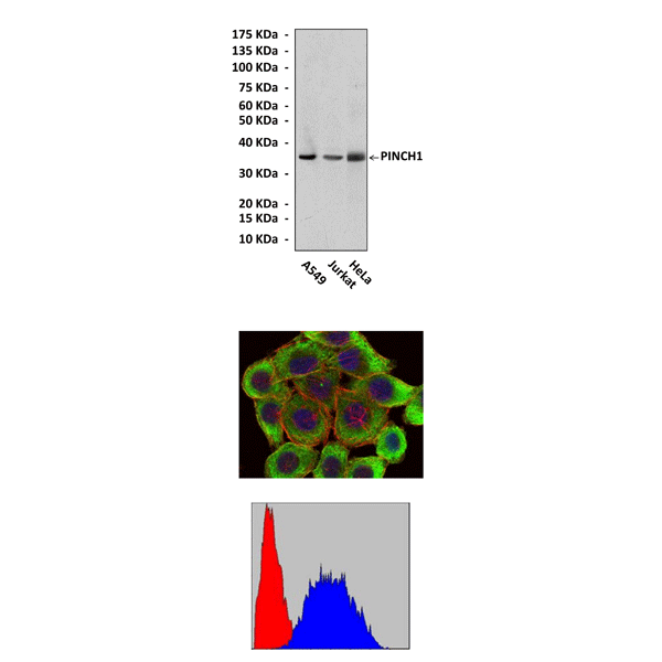
Applications & Suggested starting dilutions:* | WB 1:1000 IP n/d IHC n/d ICC 1:50 - 1:200 FACS 1:50 - 1:200 |
Predicted Molecular Weight of protein: | 37 kDa |
Specificity/Sensitivity: | Detects PINCH1 proteins in various cell lysate. |
Storage: | Store at -20°C, 4°C for frequent use. Avoid repeated freeze-thaw cycles. |
*Optimal working dilutions must be determined by end user.
Документы
Информация представлена исключительно в ознакомительных целях и ни при каких условиях не является публичной офертой


