

Anti-Syt1: Mouse Synaptotagmin 1 Antibody |
 |
BACKGROUND Synaptotagmins constitute a family of membrane-trafficking proteins that are characterized by an N-terminal TMR, a variable linker, and two C-terminal C2-domains. Synaptotagmin 1 (Syt 1) was identified as p65 in a monoclonal antibody screen for synaptic proteins and proposed as a potential Ca2+ sensor for regulated exocytosis. Synaptotagmin 1 is thought to convey the calcium signal onto the core secretory machinery. Its cytosolic portion mainly consists of two C2 domains, which upon calcium binding are enabled to bind to acidic lipid bilayers. It was suggested that synaptotagmin 1 functions by binding in a trans configuration whereby the C2A domain binds to the synaptic vesicle and the C2B binds to the PI(4,5)P2-enriched plasma membrane.1 Twelve additional synaptotagmins were subsequently discovered. Extensive work showed that Syts 1 and 2 likely function as Ca2+ sensors in synaptic vesicle exocytosis. The most abundant of these “other” synaptotagmins, Syts 3 and 7, are localized on the plasma membrane opposite to synaptic vesicles and exhibit distinct Ca2+ affinities, suggesting that plasma membrane and vesicular synaptotagmins may function as complementary Ca2+ sensors in exocytosis with a hierarchy of Ca2+ affinities. Thus, members from the synaptotagmin family are believed to underlie at least in part the distinct Ca2+ dependencies of different forms of regulated secretion. In neurons, synaptotagmin 1 (Syt1) is a Ca2+ sensor that interacts with SNAREs and membranes to mediate synaptic vesicle fusion, triggering synchronous neurotransmission. Synaptotagmin-1 mediates fast neurotransmitter release at the hippocampus, while fast release is triggered in other brain regions by this same isoform or by the closely related synaptotagmins-2 or -9. In contrast, insulin secretion in pancreatic β cells depends on synaptotagmin-7, and acrosomal exocytosis in sperm appears to involve synaptotagmin-6. In PC12 cells, Ca2+-dependent secretion is mediated by synaptotagmins-1 and -9, whereas chromaffin cells use synaptotagmins-1 and –7 as Ca2+ sensors. Functional differentiation among synaptotagmins is expected to arise primarily from differences in the properties of the two C2 domains that form most of their cytoplasmic regions (referred to as C2A and C2B domains).3 In addition, synaptotagmins also function in endocytosis. Syt 1 regulates endocytosis of the acetylcholine receptor, a process that involves the AP2 complex before membrane internalization. Syt 7 appears to have a role in membrane sealing and lysosomal trafficking.4
REFERENCES
1. Radhakrishnan, A. et al: J. Biol. Chem. 285:25749-60, 2009
2. Martens, S. et al: Science 316:1205-8, 2007
3. Xue, M. et al: PLoS ONE 5:e12544, 2010
4. Musch, M.W. et al: Am. J. Physiol. Gastrointest. Liver Physiol. 298:G203-11, 2010
2. Martens, S. et al: Science 316:1205-8, 2007
3. Xue, M. et al: PLoS ONE 5:e12544, 2010
4. Musch, M.W. et al: Am. J. Physiol. Gastrointest. Liver Physiol. 298:G203-11, 2010
Products are for research use only. They are not intended for human, animal, or diagnostic applications.
Параметры
Cat.No.: | CP10345 |
Antigen: | Raised against recombinant human Syt1 fragments expressed in E. coli. |
Isotype: | Mouse IgG1 |
Species & predicted species cross- reactivity ( ): | Human, Mouse, Rat |
Applications & Suggested starting dilutions:* | WB 1:1000 IP n/d IHC n/d ICC n/d FACS n/d |
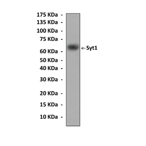
Predicted Molecular Weight of protein: | 65 kDa |
Specificity/Sensitivity: | Detects Syt1 proteins without cross-reactivity with other family members. |
Storage: | Store at -20°C, 4°C for frequent use. Avoid repeated freeze-thaw cycles. |
*Optimal working dilutions must be determined by end user.
Документы

Информация представлена исключительно в ознакомительных целях и ни при каких условиях не является публичной офертой


